Slipped Vertebra (Spondylolisthesis)

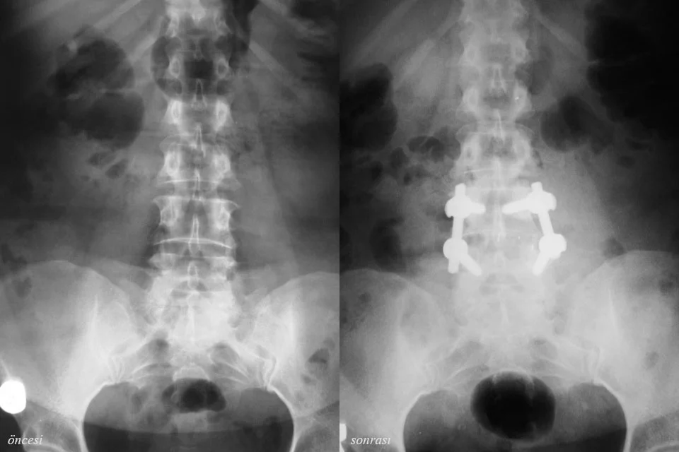
The spine is made up of a series of connected bones called vertebrae. In 5% of the adult population, a fracture can occur in the region of bone (the pars, in a condition called spondylolysis) that joins the upper and lower joints of the lowest vertebrae of the lower back. These fractures may fail to heal and can cause chronic low back pain. Sometimes the upper vertebrae can slip forward relative to the vertebra below. This condition is known as a "slipped vertebra," or as "spondylolisthesis" in the medical literature. A slipped vertebra can often be accompanied by spinal stenosis (a narrow spinal canal). The clinical signs of a slipped vertebra are low back pain, pain behind the hip joint, spasm in the lower back muscles, and, less commonly, numbness and pain in the legs and loss of strength in the feet. The pain usually increases with standing, walking, and other activities, and decreases with rest. The diagnosis is made with X-ray, bone scintigraphy, CT, and MRI.
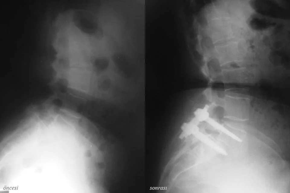
The first-line treatment consists of the non-surgical methods we call conservative treatment. Conservative methods include bed rest, painkillers and anti-inflammatory medications, temporary use of a brace, and physical therapy. If, along with low back pain, there is leg pain and numbness due to nerve compression, epidural injections may be given. In patients whose complaints do not resolve with conservative treatment methods, surgical treatment may be necessary. In spondylolysis, the fractured area is repaired and spinal fusion is performed. In spondylolisthesis, the gold-standard surgical treatment is spinal fusion. At the same time, if there is pressure on the spinal cord, the compressing structures are cleared in order to relieve it.