
Scoliosis in Children
What is scoliosis?
Viewed from the side, the spine has normal curves: a convex curve in the upper back (kyphosis) and a concave curve in the lower back (lordosis). By scoliosis we mean a sideways curvature of the spine combined with rotation. Seen from the front or the back, the trunk appears to lean to one side. Hunchback, or kyphosis, is an increase in the convex curve of the upper back.
What are the typical signs of scoliosis?
The typical signs of scoliosis are:
- A difference in the height of the two shoulders
- One shoulder blade being more prominent than the other
- One hip sitting higher or appearing more prominent than the other
- Asymmetry developing in the waist area
- One side of the back appearing higher than the other when the patient bends forward
Is scoliosis common? What causes it?
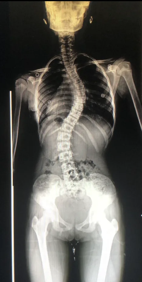
Scoliosis affects 2-4% of the population and particularly affects adolescent girls. In Türkiye there are roughly 200,000-800,000 girls and women with scoliosis. If scoliosis is mild it may not be noticeable from the outside and can only be identified through radiological examination. Advanced scoliosis is easily noticed because it causes significant cosmetic problems in the trunk.
A great deal of research is being carried out into the cause of idiopathic scoliosis, the form we encounter most often, whose cause is unknown. In the past the pathology was thought to lie in the coronal (front-to-back) plane; however, recent studies have found that sagittal (side-view) changes also accompany the problem. Experimental studies have shown that in scoliosis the process begins with a deformity in the sagittal plane, which causes rotation, after which changes develop in the coronal plane. Viewed from above, from the front and from the side, scoliosis is a complex three-dimensional deformity. It arises as a result of growth asymmetry.
Habits such as carrying a heavy bag, or always carrying a bag on the same side, play no part in the development of scoliosis.
How do we make the diagnosis?
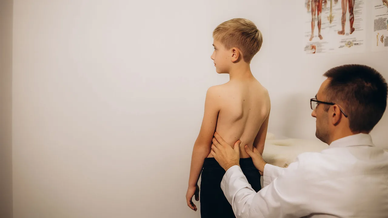
As with every condition, early diagnosis matters in scoliosis. The Adams forward-bend test, which families can easily perform themselves, allows scoliosis to be suspected. We ask the child to bend forward and look from behind: a difference in height between the two sides of the rib cage or the waist, or bony prominences along the back that do not follow a straight line, raise our suspicion of scoliosis.
As physicians, we first examine children referred to us in detail. We have a measuring device called a scoliometer. We ask the child to bend forward, place the device over the spine and take a measurement. If we obtain a value above 5 degrees, we always take an X-ray of the child. A spine that rotates and curves more than 10 degrees to the right or left on the X-ray confirms the diagnosis of scoliosis.
Do exercise and Pilates have a place in treatment?
The role of exercise and Pilates in the treatment of scoliosis is still debated. In the 1970s, bracing and surgery were at the forefront of scoliosis treatment. Between 1980 and 1990, exercise programmes for scoliosis developed by Katharina Schroth in Germany became popular. Core and Pilates exercises are beneficial particularly in postural disorders, kyphosis and scoliosis: Pilates strengthens the core muscles, increases flexibility and improves balance.
One study of the Schroth method reported that exercise had a significant effect in reducing the Cobb angle in adolescents with scoliosis of between 10 and 30 degrees. Another study of the Schroth method, published in 2024, found a difference of only 2.5 degrees between those who exercised and those who did not. More research is needed before a firm conclusion can be reached.
When is brace treatment used?
Brace treatment may be offered for scoliosis in patients who are still growing and have not completed skeletal maturity. In adolescent idiopathic scoliosis we apply brace treatment in growing children with curves between 20 and 45 degrees. Among non-surgical treatments, bracing is the most effective method for improving scoliosis and preventing progression.
A brace is made to measure for each patient's body. In recent years we have been using computer-assisted braces. The brace is made by orthotics and prosthetics specialists. The patient's waist and chest circumference and their anteroposterior and lateral diameters are measured, and a body scan is performed with a special scanning tablet. These measurements and the patient's X-rays are entered into a dedicated computer program. A thoracolumbosacral orthosis made of thin plastic is then produced by a special machine. The brace works on the three-point pressure principle.
For brace treatment to succeed, the following are required: use in mild to moderate curves, regular follow-up, a brace made correctly for the patient, a compliant patient and a supportive family, and wear of at least 20 hours a day.
How is scoliosis treated surgically?
In children who have completed growth (menstruating for two years), curves exceeding 50 degrees in the upper back and 40 degrees in the lower back require surgery. What happens if adolescents with curves above 50 degrees are not operated on? Because their curves will increase by between 1.1 and 2.5 degrees per year, 40 years later their quality of life will be poor, they will experience shortness of breath, they will have pain at an early age, they will have difficulties in social settings, and psychological problems will be more common.
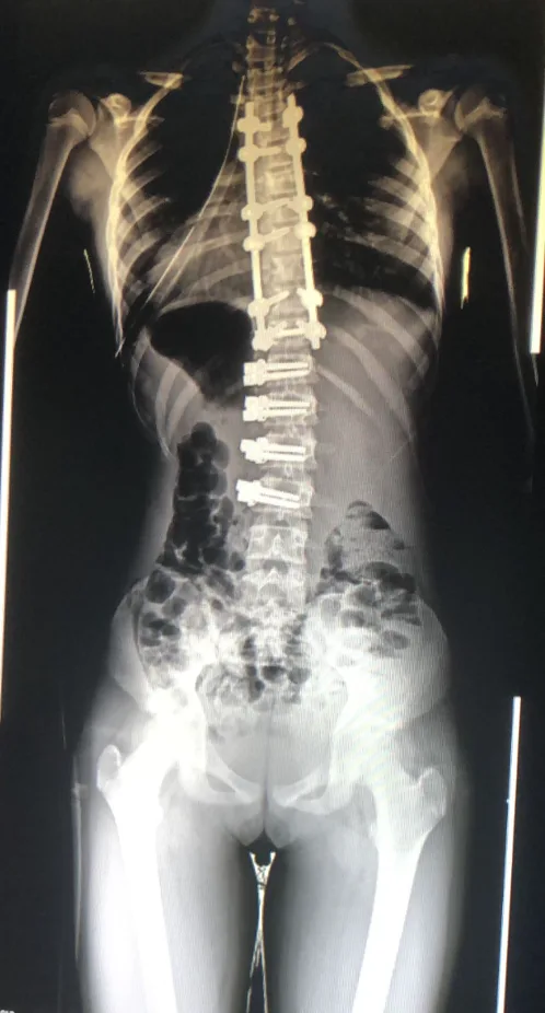
The most frequently applied method at this stage is placing screws into the vertebrae from the back, connecting them to one another with a pair of rods, performing the correction and carrying out a fusion procedure. In experienced hands the correction rate is currently around 70-80%. This means that a small scoliosis remains on the X-ray, while the child looks normal from the outside.
Vertebral body tethering (VBT)
Another newer method we use is VBT, also known as spinal tethering. It is applied in carefully selected children around the age of 10 and corrects scoliosis while leaving the vertebrae mobile, without performing a fusion.
Fusion remains the gold standard in the surgical treatment of adolescent idiopathic scoliosis. In recent years the tethering (VBT) method has become popular. Tethering is used in growing children when the curve exceeds 35 degrees. Screws and a cord are applied to the convex side of the curve, slowing growth on that side and allowing the scoliosis to correct. However, looking at the mid-term results of the tethering method in recent years, we see complications develop in one in four cases. For this reason we recommend VBT particularly in selected cases.
How is scoliosis treated in children under 10?
Scoliosis that develops before the age of 10 is called early-onset scoliosis. It can develop for a variety of reasons: most commonly we see idiopathic, congenital and neuromuscular scoliosis; less frequently we see syndromic scoliosis, scoliosis associated with connective tissue disorders such as Marfan syndrome, scoliosis related to dwarfism, and scoliosis developing after chest surgery.
For scoliosis beginning under the age of 5, early detection, determining the type and starting treatment early are important. The treatment of congenital and idiopathic scoliosis differs considerably. Assessment of infantile scoliosis detected between the ages of 0 and 3 is important; in these patients, special measurements allow us to form an opinion about whether the curve will progress.
Most of the scoliosis we see within the first year of life develops as a result of the baby being compressed in the womb, and most of these curves resolve on their own. In congenital scoliosis, however, if no intervention is made the child's lungs and heart will be adversely affected, with potentially fatal consequences. If congenital scoliosis is due to a hemivertebra or a fused vertebra, these patients need to be treated after the age of 2 without leaving it too late. Otherwise we may encounter very advanced scoliosis in adolescence or adulthood.
In children under 10 we use methods such as bracing, casting, fusion and hemi-fusion (hemiepiphysiodesis) procedures, removal of a hemivertebra, growth-guiding vertebral body tethering procedures, growth-friendly surgery, and lengthening procedures with magnetic rods.