
Microsurgery in Spinal Conditions
How is a herniated disc treated?
A lumbar disc herniation occurs when the disc between the lumbar vertebrae protrudes outward and presses on, and chemically irritates, the nerves running to the legs and the spinal cord. Disc herniation is usually seen in the lowest two discs of the lumbar spine, in the lower vertebrae of the lower back.
The classic signs of a herniated disc are pain, numbness, tingling or weakness in the legs. This painful condition is called "sciatic pain".
Non-surgical treatment first
Conservative treatments used in disc herniation are short-term rest, anti-inflammatory drugs, painkillers, muscle relaxants, physical therapy, McKenzie exercises, short-term bracing, or transforaminal and caudal epidural steroid (cortisone) injections. In 80% of patients these treatments produce results within the first three months and the patient improves.
Where these treatments do not provide relief, where marked weakness develops in standing, and in patients who develop urinary or faecal incontinence, surgery is always required. The aim is to prevent the herniated disc from irritating the nerves through pressure and thereby causing symptoms such as pain and loss of strength.
Microdiscectomy
The most frequently used methods are microdiscectomy and endoscopic discectomy. The aim is to remove the fragment of disc causing the compression.
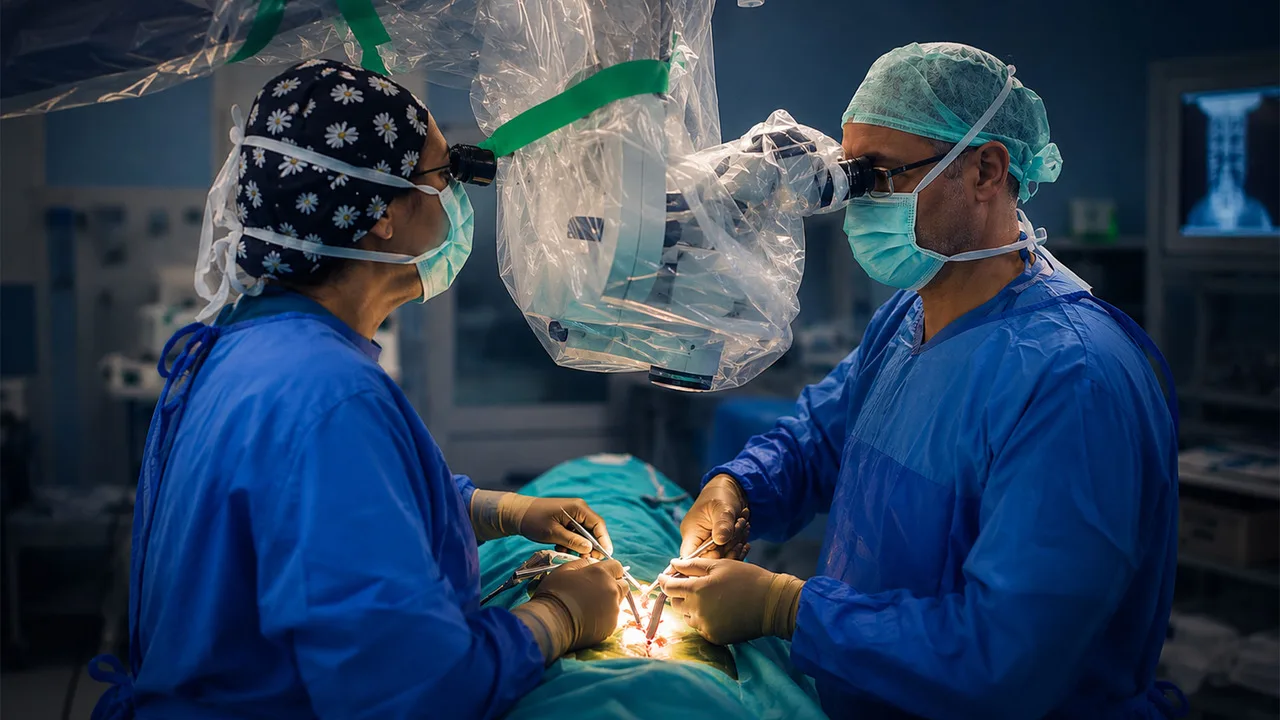
Microdiscectomy is performed through a 2 cm incision. Under the microscope, the compressing disc fragment is reached and removed. The patient is mobilised after 6 hours, mobilised with a brace, and discharged one day later.
Endoscopic discectomy
There are two techniques of endoscopic discectomy: unilateral biportal endoscopy (UBE) and the uniportal endoscopic method. In the UBE procedure, tubes are inserted through two 0.5 cm incisions in the lower back. Using a camera passed through the tube, the herniation is reached in a fluid medium and removed. Because only a small amount of intervention is made to muscle and bone tissue with this method, post-operative pain is also low. The patient can stand and walk after 6 hours.
Recovery after surgery
After endoscopic or microsurgical disc surgery, the patient returns to work within 10 days. Return to sport takes 2-3 months. The reason is that a tear forms at the back of the disc as the herniation develops, and healing of this area with scar tissue takes 2-3 months. If the patient returns to sport earlier, the likelihood of the herniation recurring is higher.
In studies comparing microsurgery and endoscopy in the literature, there is no difference between them in recurrence rates. The herniation may recur in 8-13% of cases after surgery.
How is lumbar spinal stenosis treated?
We see spinal stenosis more frequently in older patients; sometimes we also see congenital stenosis at a younger age, in the 40s and 50s.
While walking difficulties in older people are usually due to lower-back, hip and knee problems, stenosis in the lumbar region causes numbness that begins in the hips and extends into the legs, forcing the patient to stop after walking for a while.
Conservative treatment is used first: medications such as muscle relaxants, NSAIDs, gabapentin, pregabalin and cortisone (intramuscular injection). If symptoms do not resolve, epidural injections may be given. In patients who do not benefit from conservative treatment, surgical treatment is decided upon.
How is spinal stenosis treated surgically?
On investigation, the levels where the stenosis is severe are the ones producing the symptoms. We treat only those levels; there is no need to intervene at every level.
In surgery, access is gained from one side and the spinal canal is fully decompressed. We perform this procedure using either microsurgery or endoscopy. During surgery, under the microscope or with the endoscope, the thickened ligamentum flavum compressing the canal must be removed, along with the bony spurs arising from the arthritic joint.
Following this procedure the patient's symptoms resolve within a short time. The patient is mobilised and walked on the first day and is usually discharged within 1 or 2 days. A brace is used for 1 month. The patient returns to work after 2 weeks. Patients are kept away from heavy work for 3 months.